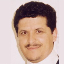
Habib Zaidi
Geneva University Hospital, Switzerland
Quantitative imaging biomarkers
in the era of precision medicine
Abstract
Early diagnosis and therapy increasingly operate at the cellular, molecular or even at the genetic level. As diagnostic techniques transition from the systems to the molecular level, the role of multimodality molecular imaging becomes increasingly important. Positron emission tomography (PET), x-ray computed tomography (CT) and magnetic resonance imaging (MRI) and their combinations (PET/CT and PET/MRI) provide powerful multimodality techniques for in vivo imaging. Quantitative image analysis has deep roots in the usage of molecular imaging in clinical and research settings to address a wide variety of diseases. It has been extensively employed to assess molecular and physiological biomarkers in vivo in healthy and disease states, in oncology, cardiology, neurology, and psychiatry. This talk reflects the tremendous increase in multimodality molecular imaging as both clinical and research imaging modalities in the past decade. An overview of advanced medial image instrumentation technologies and PET image quantification and related image processing issues with special emphasis on radiomics analysis will be presented. This talk aims to bring the biomedical image processing community a review on the state-of-the-art algorithms used and under development for accurate quantitative analysis in multimodality and multiparametric molecular imaging and their validation mainly from the developer’s perspective. It will inform the audience about a series of advanced development recently carried out at the PET instrumentation & Neuroimaging Lab of Geneva University Hospital and other active research groups. Current and prospective future applications of quantitative molecular imaging are also addressed especially its use prior to therapy for dose distribution modeling and optimization of treatment volumes in external radiation therapy and patient-specific 3D dosimetry in targeted therapy towards the concept of image-guided radiation therapy. In this regard, the promising role of artificial intelligence (AI), in particular deep learning approaches, will be emphasized. To this end, example applications of deep learning in five generic fields of multimodality medical image analysis, including imaging instrumentation design, image denoising (low-dose imaging), image reconstruction quantification and segmentation, radiation dosimetry and computer-aided diagnosis and outcome prediction will be discussed. Future opportunities and the challenges facing the adoption of deep learning approaches and their role in molecular imaging research are also addressed.
Speaker’s Bio
Habib Zaidi is Chief physicist and head of the PET Instrumentation & Neuroimaging Laboratory at Geneva University Hospital and full Professor at the medical school of Geneva University. He is also a Professor of Medical Physics at the University of Groningen (Netherlands), Adjunct Professor of Medical Physics and Molecular Imaging at the University of Southern Denmark, Adjunct Professor of Medical Physics at Shahid Beheshti University visiting Professor at Tehran University of Medical Sciences and Distinguished Adjunct Professor at King Abdulaziz University, KSA. He is actively involved in developing imaging solutions for cutting-edge interdisciplinary biomedical research and clinical diagnosis in addition to lecturing undergraduate and postgraduate courses on medical physics and medical imaging. His research is supported by the EEC, Swiss National Foundation, EEC, private foundations and industry (Total 8.8 M US$) and centres on hybrid imaging instrumentation (PET/CT and PET/MRI), deep learning for various imaging applications, modelling medical imaging systems using the Monte Carlo method, development of computational anatomical models and radiation dosimetry, image reconstruction, quantification and kinetic modelling techniques in emission tomography as well as statistical image analysis, and more recently on novel design of dedicated PET and PET/MRI scanners. He was guest editor for 13 special issues of peer-reviewed journals dedicated to Medical Image Segmentation, PET Instrumentation and Novel Quantitative Techniques, Computational Anthropomorphic Anatomical Models, Respiratory and Cardiac Gating in PET Imaging, Evolving medical imaging techniques, Trends in PET quantification (2 parts), PET/MRI Instrumentation and Quantitative Procedures and Clinical Applications, Nuclear Medicine Physics & Instrumentation, and Artificial Intelligence and serves as founding Editor-in-Chief (scientific) of the British Journal of Radiology (BJR)|Open, Deputy Editor for Medical Physics, and Associate Editor or member of the editorial board of the Journal of Nuclear Cardilogy, Physica Medica, International Journal of Imaging Systems and Technology, Clinical and Translational Imaging, American Journal of Nuclear Medicine and Molecular Imaging, Brain Imaging Methods (Frontiers in Neuroscience & Neurology), Cancer Translational Medicine and the IAEA AMPLE Platform in Medical Physics. He has been elevated to the grade of fellow of the IEEE, AIMBE, AAPM, IOMP, AAIA and the BIR and was elected liaison representative of the International Organization for Medical Physics (IOMP) to the World Health Organization (WHO) and Chair of Subcommittee on Part 1 Examination of the International Medical Physics Certification Board (IMPCB) and the Imaging Physics Committee of the AAPM in addition to being affiliated to several International medical physics and nuclear medicine organisations. He is developer of physics webbased instructional modules for the RSNA and Editor of IPEM’s Nuclear Medicine web-based instructional modules. He is involved in the evaluation of research proposals for European and International granting organisations and participates in the organisation of International symposia and conferences. His academic accomplishments in the area of quantitative PET imaging have been well recognized by his peers and by the medical imaging communi ty at large since he is a recipient of many awards and distinctions among which the prestigious 2003 Bruce Hasegawa Young Investigator Medical Imaging Science Award given by the Nuclear Medical and Imaging Sciences Technical Committee of the IEEE, the 2004 Mark Tetalman Memorial Award given by the Society of Nuclear Medicine, the 2007 Young Scientist Prize in Biological Physics given by the International Union of Pure and Applied Physics (IUPAP), the prestigious (100’000$) 2010 kuwait Prize of Applied sciences (known as the Middle Eastern Nobel Prize) given by the Kuwait Foundation for the Advancement of Sciences (KFAS) for “outstanding accomplishments in Biomedical technology”, the 2013 John S. Laughlin Young Scientist Award given by the AAPM, the 2013 Vikram Sarabhai Oration Award given by the Society of Nuclear Medicine, India (SNMI), the 2015 Sir Godfrey Hounsfield Award given by the British Institute of Radiology (BIR), the 2017 IBA-Europhysics Prize given by the European Physical Society (EPS) and the 2019 Khwarizmi International Award given by the Iranian Research Organization for Science and Technology (IROST). Prof. Zaidi has been an invited speaker of over 160 keynote lectures and talks at an International level, has authored over 850+ publications (he is the senior or first author in a majority of these publications), including 375 peer-reviewed journal articles in high ranking journals, most of them in Q1/D1 of their categories (h-index=73, >191’0+ citations | Google scholar), 425 conference proceedings and 42 book chapters and is the editor of four textbooks on Therapeutic Applications of Monte Carlo Calculations in Nuclear Medicine (2 Editions), Quantitative Analysis in Nuclear Medicine Imaging, Molecular Imaging of Small Animals and Computational anatomical animal models.
